Friday, February 20, 2009

Better Late Than Never: AGAIN.......and AGAIN.....

This week’s lab began with our group finishing the plating of our twenty-six strains on optimal media containing starch. This test will be used to further characterize our strains. The next task involved scoring the growth of our twenty-six strains in liquid culture media. Our group then preformed colony counts of the irradiated Little Red Hill soil sample. The decrease in number of colonies with increase of dilution demonstrated good dilution technique.
The plates also showed great diversity across the media we used. This maybe the result of the media facilitating the growth of certain organisms while inhibiting the growth of other organisms.
Our group took 10 micro liters from each of the irradiated soil dilutions and plated them on new MA, PCA, 1/10 PCA, and 1/100 PCA media. Our group then selected 10 suspected organisms from the irradiated plates and plating them on new media. We parafilmed the plates and placed them in the incubator. From the results of the colony counts we determined the cfu/g for our irradiated soil samples.
THERE WAS A NICE EXCEL TABLE BUT THIS BLOG CAN COPE WITH TABLES.... the tables are at AirSet
We determined the percentage of survival by comparing the cfu/g of the irradiated soil counts over the cfu/g of the non-irradiate d soil counts.

THERE WAS A NICE EXCEL TABLE BUT THIS BLOG CAN COPE WITH TABLES.... the tables are at AirSet

It can be noted that the radiation significantly reduced the number of organisms in all cases with the exception of one of the Gobi samples. This lead to a class discussion of some of the possible reasons why the survivorship increased in the irradiated sample.
Our second lab day began with scoring the temperature variation plates of our twenty-six strains (10, 25, 30, 37, and 42 degrees Celsius) for growth and recording the data into our master chart. It should be noted due to a malfunction of the refrigerator, our 10 degree Celsius results may have been altered. It is also noted that 1/10 PCA media was contaminated, so three of our samples were bad. We then scored the growth of the twenty six strains, compared to the 25 degree Celsius plates, on media of varying NaCl concentrations (1, 3, 6, and 9%) and recorded that data into our master chart. For some reason our group was missing our strain 39 NaCl plates. There was significant variation of growth and diversity between the various temperatures and NaCl concentrations. This may be due to the fact that those variations inhibited or simulated the growth of certain organisms. Our group then streaked the twenty-six strains from the 25 degree Celsius plates to new optimal media to use as our new stock culture plates. For the samples that were contaminated we used our original stock vials to make new stock culture plates. Our class for the week ended with a group discussion of the upcoming midterm.

Don’t used borrowed media.............

This was a busy week on Monday we counted our irradiated soil plates. We then took theses counts and used them to determine the percent survival of organisms compared to the un-irradiated plates that we counted earlier. For our organisms the higher percent survival and diversity was on the lower nutrient plates this is most likely due to the resemblance of the availability of nutrients on these plates to the natural environments of the organisms. Our Isolates did not like salt after being irradiated nothing grew on them at all. We used the irradiated soil plus media enrichment broths to streak plates to see what grows along with picking 10 colonies from the irradiated plates that most resembled our current isolates to do further test too. There is a graph in a paper given to us by Dr. Rainey that has a graph of the survival percentages after different doses or ionizing radiation. On this graph it shows that with a 5kGy dose which is what we gave our soil sample the survival percentage is 20% our data gives a lower surviving percentage anywhere from 0-18%.

Tuesday was all about grading plates and a little Midterm review. For temperature plates it was surprising the range of growth. The majority of the isolates grew at just about every temperature. What was surprising to me was that at the higher temperatures some organisms changed there pigmentation. I believe this could be temperature affecting the enzymes that typically make these pigments. For salt the range of optimal growth tended to be 0-3% with the few isolates doing well on 6% and only a single one doing anywhere near decent growth on 9%. We received variable results from the isolates that typically grow on MA some did better on R2A with 3% salt than they did on MA alone. Also it was noticed that just because it likes a little salt doesn’t necessarily meant it will grow better at higher salt concentrations.

The most probable reason for the contaminated 1/10th PCA plates was the media was already contaminated before use. This can be deduced by the imbedded colonies in the media.
The Plate picture story is that Salt was not liked at all after irradiation. High nutrients was not favored for numbers or diversity after irradiation. 1/10th PCA was the best to bring around the highest numbers and diversity with 1/100th having high numbers also but not as high diversity.

N97 irradiated soil 10-1 plated on 4 different media and incubated for 20days at 25C:
MA:

PCA:

1/10 PCA:

1/100 PCA:

wAtCh OUT

On Monday, Feb. 16, we had a LOT to do. We counted our irradiated soil plates and calculated the cfu/g, the percent survival and 5kGy, completed streaking our starch plates, and picked 10 more colonies, 11-20, and took pictures of them. We also checked for growth in our liquid cultures, scored them, and streaked them out using 10 microliters. Before we streaked the plates we had to vortex the tubes to insure that the everything was thoroughly mixed. When we counted our irradiated plates our (-1) plates were the most countable. Our (-2 and -3) plates had very little to no growth. They also had some contamination. I find it very interesting that we had very little growth on our (-2 and -3) plates at 5kGy. That makes me think that at the higher dosages we won't have any survival on those plates. It should also be noted that most of our growth on our (-1) plates occurred on 1/10 PCA and 1/100 PCA. To me, this seems like the organisms don't require a lot of nutrients to grow. The percent survival for the 1/10 PCA and the 1/100PCA plates were only 8.4% and 16.4%, respectively. I find it very interesting that Golbi had such high percentages of growth on all of the media. In addition, when we scored our liquid cultures we noticed that every tube had growth. So, we scored them from 1-4+. We took out 4 totally different tubes, one that would be representative of 1-4+. By having something to compare the other tubes to made it a lot easier. Most of our tubes had 2+ growth. Next, when we chose our 10 colonies to streak out, we chose 6 black and 4 pink. I don't know for the other groups, but for us, we had tons of black colonies on our plates. In looking at our irradiated soil plates you will see that most of our growth did in fact occur on our 1/10 PCA and 1/100 PCA plates. The other media had very little to no growth.

LRH - growth on 1/10 PCA after irradiation: 
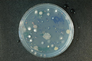
LRH - growth on 1/100 PCA after irradiation:
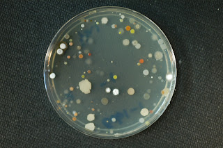
LRH - growth on PCA after irradiation:
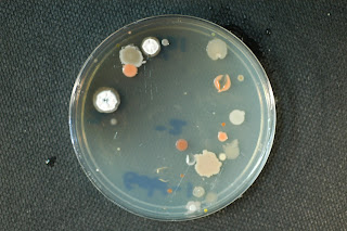
LRH - growth on MA after irradiation:


On Wednesday, Feb. 18, we had a LOT to do AGAIN. HaHaHaaa!! I arrived a few minutes late (16 to be exact) because I couldn't find my key. The entire time it had been in my purse!! But, on a heavier note, when I arrived I found my partners looking at our temperature and NaCl plates. We took note of the growth, 1-4+, and the color of the colonies on the plates. In looking at our plates we noticed that we had lots of growth across the board. Every temperature had lots of growth. It was very surprising that we had growth the on the plates stored in the 10 degrees centigrade incubator. The organisms that survived on these plates obviously come from or have some resistance to cold environments. In addition, our NaCl plates had lots of growth too. However, as the NaCl was increased (1,3,6,9) we noticed that growth decreased. There were some that did grow very well. I think that these organisms are from the Modestabacter genera. I'm not sure so someone if you know please correct me on this. We don't have results for our 1/10 PCA plates because the incubator went out for a while and went to 25 degrees centigrade. Maybe, JUST MAYBE, someone should buy a more expensive incubator and not be so CHEAP!! That was a JOKE!! HaHAahaha… Later that day, we made stock cultures from 23 , 25 degrees centigrade plates, and 3 (the 1/10 PCA) from the cold room.

I met the Duggarts yesterday in free speech alley with all 17 kids...

On 2/16/09 we did plate counts for the radiated soil samples that were plated on the various media.. We used these counts to calculate the colony forming units per gram of soil. Also, we compared our data from the unradiated soil samples and calculated the percent survival.

Percent Survival= (Unradiated cfu/g / Radiated cfu/g) x 100
MA=6.87%
PCA=41.78%
1/100 PCA=227.6%
1/10 PCA=74.8%
*MA had the largest reduction
*1/100 PCA is a freak of nature (no, not really! I explain it below)


We photographed the one of the -1 dilution plates from each media and one plate from each dilution of 1/10 PCA. Most of the data from the three soil samples (Golbi-1, LRH, and N97-2) agreed with our hypothesis that radiation would lower the plate counts. However, the Golbi-1 radiated soil sample differed from this pattern. Although the cfu/g from the radiated soil sample descreased on PCA and 1/10 PCA, the % survival was higher compared to the other groups with 41.8% and 74.8% respectively. On the other hand, the cfu/g from the radiated soil sample on the 1/100 PCA differed greatly from the results of the other media and soil samples. Our cfu/g increased over 100% as compared to the unradiated soil sample. At first, I assumed that we must have made a mistake. However, upon a little research and brainstorming I have formed a couple of hypothesis to explain this mysterious data. One possibility that I shared in class is that a competitor with the radiation resistant strains was eliminated upon radiation; therefore, the cfu/g would increase. Another possible explaination that I recently came across comes from an article in Nature. In this article, it talks about the advantages of high intracellular manganese and low intracellular iron in radiation resistant organisms. The high intracellular manganese reacts with free radicals produced during a cells metabolisms. Therefore, the bacteria cell is healthier overall. This hypothesis was tested by lowering the intracellular manganese in radiation resistant organisms in which they became more sensitive to DNA damage and cell death. This research is not only beneficial to our study on radiation resistant microorganisms, but it could also be a great tool in minimizes radiation damage to healthy tissue in cancer patients. By increasing the healthy tissues intracellular manganese and not adjusting the concentration in the target cancer cells, side effects from radiation could be greatly reduced. Anyways, back to bacteria!
Our 78 strains were also streaked on starch containing media. The plate was streaked in order to make somewhat of a lawn of cells so that we can clearly visualize clearings if the cells do in fact metabolize the starch. Someone comment on this and tell me if I’m right in saying that we are testing for amylase production? If not, what enzyme are we testing for? The culture were incubated at 25 C.
Next, we recorded the growth in the liquid culture of our 26 stains which were inoculated in the broth on 1/26/09. We measured this growth by vortex the broth and visualizing the turbidity. All of our strains grew in the liquid media. However, some had more growth than others. I am e-mailing a table of the data we recorded from this experiment. Strain #68 was not done because it was not inoculated on 1/26/09 due to contamination. We made a liquid broth and inoculated it with strain #68 on Monday, 2/16/09, and will check it in 20 days. The growth was rate on a scale of 1-4 with 4 being the greatest amount of growth and 1 being little growth.
Dilution plates (-1, -2, -3) were made of the radiated strains. 10 ul were streaked on each plate. Only one plate was streaked per dilution.
From the plate counts on the radiated soil samples, 10 colonies were selected as possible Modestobacter suspects. They are denoted GB11-GB20. I am really excited because we actually got a black colony. I am adding these new isolates to the Modestobacter suspect chart I made from the unradiated soil sample.
Wednesday was a hectic day. We recorded the growth from the temperature and [NaCl] plates. This should have been fairly simple, but it ended up being a really big headache because we miss placed some of the plates. Our observations of these plates is including in the spread sheet on airset. I noticed that not only does temperature and [NaCl] affect colony growth, but it also affects the color. The color is also recorded in the spreadsheet on airset. Our data is recorded using a scale of (0-4+) with zero being no growth and 4+ being the most growth. We have finally overcome the great strain #56 anomaly. #56 hates MA! Why on earth we thought that it grew best on that on our first day of class is beyond me. #56 grows best on PCA. For now on MA and 56 will have nothing to do with each other. Anyhow, 59 and 69 had to be restreaked for the temperature plates and observations will be recorded at a later date. This is due to contamination on their temperature plates. Also, 68 was not originally done on both the temperature and salt plates because the original culture was contaminated. Sean Michael made new plates for temperature and salts for 68. Today was a disaster.

Irradiated Golbi -1 dilution 1/100 PCA plate:

Blog............

This week in class we counted our irradiated soil plates, calculated the colony forming unites per gram of soil, calculated the percent survival rate, took pictures, and chose ten colonies from those plates to restreak onto fresh plates. We had significantly less growth on our irradiated soil plated that were exposed to 5 kGy radiation compared to our unirradiated plates. On our nonirradiated plates we had the most growth and diversity on 1/10 and 1/100 PCA with significant growth up to 10-4. The 1/100 PCA had the highest cfu/g at 2.276 x 10⁴. For our irradiated soil plates we had no growth at all on MA and on PCA we only observed growth on two of the 10-1 plates. Once again the best growth was observed on 1/10 and 1/100 PCA. The most colony forming unites per gram of soil was on the 1/10 PCA at 2.67 x 10³. There was less diversity on the irradiated plates which is to be expected since most organisms are sensitive to radiation. Most of the colonies present were pink/orange/peach (see figure below).
We had the greatest percent survival on 1/10 PCA with 18.7% and the lowest on MA with 0%. Overall radiation reduces the number of colonies present as well as the diversity.
We also observed our salt and temperature plates. Surprisingly we had multiple strains that grew very well on 10°C and a couple that grew well on 42°C. We now need to lower and raise the temperatures to conclude the temperature limits in which these organisms can survive. We were not able to record data for strains grown on 1/10 PCA due to contamination of the media. I believe the temperature at which we had the most growth present was 30°C. Not many of our organisms survived on 9% NaCl and only slight growth was observed at 6%. With the exception of a few strains, most of them grew well on both 1% and 3%, with the greatest amount of growth on 1%. In class we also finished streaking our strains onto starch agar that did not get done last week. We also observed our strains in liquid media to check for growth. Growth was present in all the liquid media.
We streaked 10µl of the irradiated liquid soil dilutions onto plates. Finally, we took pictures of our soil plates. Below you can see our 1/10PCA 10-1 plates pre and post irradiation. The colonies present on the unirradiated soil plates were more tan and cream colored while the colonies on the irradiated plate were pink and less plentiful. I believe this is due to the fact that the tan colonies were most likely inhibiting the growth of the pink colonies and once they were killed off by radiation the pink colonies which were radiation resistant were able to grow.

1/10 PCA 10-1 dilution for N97 pre-irradiation:

1/10 PCA 10-1 dilution for N97 post-irradiation (5kGy):

Week Summary

On Monday of this week, we streaked the remaining starch plates that we were unable to do the week before. Then we made observations of the growth of our strands in the liquid media. We scored them from 0 to ++++. We had growth in all of our samples ranging from + (included 59, 73, 74, 64, 65, and 78) up to ++++ (included 54, 56, 67, 69, and 70). Sample 68 was contaminated. So some of the stock culture was again placed in PCB liquid media and will be checked again in 20 days. Then we counted cultures on irradiated Gobi-1 samples, and found that MA had by far the lowest CFU/g of only 6.6x10². On the un-irradiated samples, MA had a CFU/g value of 9.7x10³, giving it only 6.9% survival. This may have occurred because most of the bacteria that require salt have a low irradiation tolerance. For the other irradiated samples we found that PCA had a CFU/g of 1.1x10³ with a 48.1% survival, 1/10 PCA had a CFU/g 7.63x10³ with a 74.8% survival, and 1/100 PCA had a CFU/g of 1.32x10^4 with a 227.6% survival. 1/100 PCA's unusual percent survival may have occurred because the irradiation inhibited the growth a bacteria that dominated the un-irradiated samples. In general, the irradiation had lower number of colonies, but with most of our samples having high survival percentages most of our bacteria have a high irradiation tolerance. Also, because 1/100 PCA had the highest CFU/g value, this may show that the irradiation resistant bacteria prefer low nutrients.

On Wednesday, we observed and recorded the results from the temperature and salt experiments with our stands. We streaked new stock cultures from the 25ºC samples. Culture #59 and #69 had contamination in the temperature. So from the second stock culture we re-streaked the temperature trials. Also, #68 had enough growth on our stock culture. So we streaked the temperature and salt trials. The results from the salt plates showed that all the bacteria can live up to 3% except for #72 and #77. Only eight of our strands were able to survive up to 9% (58, 59, 61, 62, 63, 64, 73, and 74). Overall, most are the bacteria are fairly sensitive to higher concentrations of salt. The results from the temperature plates showed that all the strands except six (64, 66, 70, 71, 72, 74) could survive at temperatures as low as 10ºC. While only 55, 63, and 74, couldn't survive at 37ºC, and 53, 54, 57, 58, 60, 72, 75, 76, and 78 were all able to withstand temperatures as high as 42ºC.

An interesting observation is that #74 had a small temperature range only growing at 25ºC and 30ºC, but it grew in media that contained 9% NaCl. Being that it was orange in color, I compared it to the two Blastococcus species ( B. saxobsidens and B. jejuensis ). According to the Urzi et al 2004 paper, Blastococcus saxobsidens has a temperature growth range of 20-37ºC, and it was reported to have grown on 5% NaCl. These similar traits with further tests may reveal that #74 is Blastococcus saxobsidens.

Another Very Busy Week

This week was extremely busy and frustrating, at times, but our group accomplised a lot. On Monday we counted our irradiated soil dilution plates, completed the streaking of our starch plates, checked for and recorded growth in our liquid cultures (made from our stock cultures), selected 10 colonies from the irradiated plates that we thought could be Modestobacter and restreaked them onto new plates, and lastly we streaked 10ul from our diluted (-1,-2,-3) irradiated cultures onto media plates. When we counted our irradiated dilution plates we found that the most growth occurred on 1/100 PCA, with an average of 132 colonies per plate. We found this very interesting because this average was even higher than the average for the unirradiated 1/100 PCA plate (which had an average of 58 colonies per plate). We think this might be because one organism was inhibiting the growth of a particular species on the unirradiated plates. However, when the samples were irradiated, this inhibitor was killed, which allowed other species to flourish. The second highest growth occurred on 1/10 PCA (76.3 average colonies per plate) followed by PCA (average of 11 colonies per plate) and finally MA (6.6 colonies per plate). After counting the colonies, we took pictures of -1,-2,-3 1/10 PCA to show the effect of dilution on colony count. We also took pictures of -1 PCA, -1 MA, -1 1/10 PCA, and -1 1/100 PCA in order to show the effect of different media on colony count. Lastly, we calculated the cfu/mL for each type of media using each -1 plate and calculated the percent survival by dividing the irradiated cfu/mL by the unradiated cfu/mL. Our results are as follows:

Media Type--Unradiated cfu/mL--Irradiated cfu/mL---% Survival
---1/10 PCA---------1.02 x 10^4---------------7.63 x10^4----------74.8%--------
---1/100 PCA--------5.8 x 10^3---------------1.32 x 10^4---------227.6% (!!!!)-
----PCA------------2.633 x 10^3--------------1.1 x 10^3------------41.78%--------
-----MA-------------9.7 x 10^3-------------------666-------------------6.87%--------

From these results, we found that, in general, radiation reduces cfu/mL and that all of the groups' percentage survival ranged from 0-227.6%. From this dilution series we chose 10 colonies that we thought might be Modestobacter and attempted to isolate them by streaking them onto new plates. We chose black, peach, orange, and pink colonies from the -1 dilutions of 1/100, 1/10, PCA, and MA. We streaked two colonies per plate and incubated them at 25°C. We also finished streaking our strains onto the starch plates from our stock plates (strains: 53, 55, 56, 57, 59, 67, 70, 75, and 74). We then parafilmed these plates and incubated them at 25°C. Then, we removed our liquid stock cultures from the 25°C incubator to check for and record growth. To do this, we vortexed the tubes and then observed them for turbidity. We recorded the amount of growth using a scale of 4+ for most turbid (most growth), 1+ as the least turbid and 0 as no growth. All of our tubes did have some growth. We found a range of 1+ to 4+ when examining these tubes for growth. We did not have a liquid culture for 68 (our stock culture was contaminated on the day we made these liquid cultures), so we made a new one using PCB. We incubated this new tube and we will check it for growth in 20 days. Lastly, we removed our liquid dilution series of the irradiated samples (-1, -2, -3) and streaked 10 uL onto plates. We plated these samples onto the type of media each particular strain grows the best on. We then parafilmed and incubated them at 25°C. On Wednesday we observed and recorded growth/color of the colonies on the varying NaCl and temperature plates. Again, we used 4+ as the most growth and 1+ being little growth. These plates were compared to the 25°C temperature plates, which we used as a control. Every strain that was put under varying conditions was compared against the control plate to see if it had less or more growth (Ex: strain 53 on 1% NaCl was compared against strain 53 that was incubated at 25°C). The results our group recorded can be found on AirSet. We found that the majority of our strains preferred 1% or 3% NaCl (most had 3+ to 4+ growth). However, some of our strains were able to grow at 6% NaCl and even 9% NaCl (although the growth was very small, mostly 1+ and a few 2+). We also found that the colonies of each strain tended to change color depending on the amount of NaCl they were exposed to. For example, a colony that grew bright orange on 1% NaCl was a pastel orange on 6% NaCl. I found on the internet that this color change could be because of secondary metabolites that are produced when excess salt is present. I'm not sure if this is the case for our strains, though. For the temperature portion of the experiment, we found that most strains preferred a range from 25°C to 37°C, but there were some strains that were viable at 10°C and 42°C, as well (even some with as much as 4+ growth!!). We did not notice as much of a color change due to varying temperatures. There were a few strains that had a slight color variation correlating to temperature increase, but it was not as dramatic as color changes due to changing salt concentration.Unfortunately, the strains of 59 and 69 that were put under varying temperatures were contaminated with a fuzzy, white growth. We replated these for each temperature (using the stock plate) and we will observe these plates in 20 days. We also had to replate strain 68 for both the temperature and the salt experiment. We had to do this because this strain was not plated to begin with (the original stock plate had contamination on it. We had to restreak the stock culture onto a new plate. On the day this experiment was performed, the new stock culture had not grown yet). The last thing that we did on Wednesday was restreak our stock cultures using the plates that we had incubated at 25°C for the temperature experiment. We did this in order to keep new colonies growing, since the colonies on the original stock plates were getting old and difficult to remove. I'm sure they were also undergoing phenotype and chemotype changes due to their age.Below you can see the effects of dilution on our irradiated 1/10 PCA plates. Notice how the number of colonies drastically decreases with increasing dilution. These pictures were taken on Monday.


My Blog :-)

After this week I knew my life would never be the same! I realized that I would have to read the encyclopedia cover to cover to earn my A! On Monday we completed numerous tasks in lab. First we met in room 347 where we discussed the blogs and the importance of commenting on others' blogs! We were given our list of assignments for the day which included: completing our starch streaks from the previous class, checking the growth from our liquid cultures, vortexing and scoring our liquid cultures, streaking our liquid dilutions, counting the irradiated plates, taking pictures of our selected plates, and choosing ten to restreak. Our first task, which was to complete our starch streaks from the previous class, went fairly well. We had to streak strains: 33, 50, 52, 41, 42, 44, 47, 51 and 43 because the media that it needed to be streaked on was not available last class. We were given "strict" rules to streak the plate straight down, which is what I did. After this we checked the growth from our liquid cultures. To do this we had to vortex the tubes so that the growth which was present predominantly at the bottom of the tube would be suspended throughout the tube. We measured the growth by the amount of turbidity present. There was growth present in every tube! However, strain 50 had the most turbidity, signifying the greatest amount of growth. We then took 10 microliters of the irradiated soil dilutions and streaked it onto plates. Next we counted the irradiated plates and calculated the colony forming unit/ gram (similar to the calculations which were performed last class). The most growth was found on the 1/100 PCA media, and the least growth were found on the PCA media. On marine agar media there was an average of 17.3 colonies which is low. This could be because the organisms from this sample are not very tolerant or salt. As before, as the dilutions increased (-1 to -3) the amount of colonies decreased dramatically After counting the colonies and before doing calculations we chose ten colonies (six black and four pink) from our plates to restreak. We also calculated the percent survival (this week cfu/g divided by last week's cfu/g). The results from these calculations revealed that the not many organisms survived. The greatest percentage survival was found on the 1/100 PCA media. Thorugh this we concluded that radiation reduces population; it selects for different organisms. Those that survived were tolerant to radiation. Also, on radiated plates there are no Streptomyces and less diversity. Mostly orange and pink colonies were present. .
On Wednesday, the task load got a bit lighter. The assignments included rating NaCl and temperature plates and making new stock culture from our control plates (25ºC). The various temperatures included 10, 25, 30, 37, and 42 degrees. The different salt percentages included 1, 3, 6, and 9. For the temperature plates there most growth occurred at 30 ºC, whereas the least amount of growth was mainly at 10 ºC. This experiment has to be performed again because these plates were placed in a faulty incubator, which could account for the low growth rates at this temperature. The 1/10 PCA plates were contaminated, so we was not able to use them. The salt plates showed most growth on 1 percent NaCl and lowest growth at 9 percent NaCl. This may be because these organisms are not salt tolerant.
From the pictures below you can see that as the dilution increased less colonies were found on the plates of the irradiated soil.
LRH -1 dilution tenth strength PCA:

LRH -2 dilution tenth strength PCA:
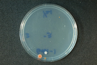
LRH -3 dilution tenth strength PCA:

Mardi Gras will always be here....

For the most part this week in lab involved recording data from experiments and tests that we set up in previous classes.

On Monday we counted the colonies on the countable plates from the serial dilution plating of soils that had been exposed to 5 kGy of gamma irradiation. We calculated the cfu/g values for each soil after 5 kGy radiation exposure and the surviving fraction. The surviving fraction was determined by comparing the cfu/g value before and after irradiation for each soil. Dr. Rainey discussed the results with us and we talked about the age of the soils, the locations of the soils sampled and the idea of competition on serial dilution plates. We found differences between the levels of survival between the soils and these data can be found at our AirSet site.
In addition we streaked out the remainder of our strains on starch agar plates (which Eugene had prepared for us). Monday was also 20 days since we inoculated our liquid cultures to determine growth in liquid media so we recorded these data. This test is a complicated one to both read and interpret. The liquid culture tubes were incubated without shaking and so after 20 days the majority of growth was in the bottom of the tubes. Before recording the growth we vortexed the tubes and scored them 0 to ++++. The majority of the strains seem to grow in liquid culture although some seem rather hydrophobic and clump.

On Monday Dr. Rainey also reaffirmed the importance of the BLOG in regards to how much effort we put into it and the fact that some in the class had not been reading or commenting on the BLOG. There is a good part of our grade involved in the BLOG and so it would be best to take it serious. It is also a wonderful way to recap on the week and learn about what we have been doing and what others in the class perspective is of what we are doing.

Recording growth at various temperatures and NaCl concentrations was the main activity on Wednesday. We found that some strains grew at all NaCl concentrations as was the case with the temperature range. A number of strains grew at the extremes tested (10C and 42C). The 10C results do in some ways have to be taken with a pinch of salt at this point as the 10C incubator went down (or up as the case was) and so they may have been at RT for a day or two before Dr. Rainey moved them to a new 10C incubator. We are going to repeat the 10C test again. For those strains growing at 10C we will try to grow then at 4C and for those growing at 42C we will try to grow them at 45 and 50C. When we set up these plates we will streak them from plates grown at 25C and not from the 10C or 42C plates as this could result in the selection or training the strain to grow at the extremes. It would seem that a lot of the strains do well at 30C. For the NaCl results we discussed the fact that those growing at the higher NaCl concentrations may have come from sample sites at which the soils have higher salt concentrations. We will compare these data between sites and with soil sample salinity in another class. The last task Wednesday was to streak a new stock plate and incubate it at 25C. Next week we will use this stock plate to streak other tests at various pH and to test the ability of the strains to degrade polysaccharides like xylan and cellulose.

At the end of class on Wednesday Dr. Rainey went over the plan for the midterm (which is on 4th March). He told us there will be 20 short questions involving a line or two of writing and 4 long questions that will have up to a page of space to write our answers. We need to know everything we have done in class and technical details of all our experiments as well as why we did what we did. We need to know what results we obtained and what they might mean and how they will be used in the overall aim of our class which is to characterize and describe new species of the genera we are working with. The BLOG and the comments will also be a valuable resource for eth midterm along with our lab notebooks. The worst part is that we need to read the papers he put up on AirSet but he did tell us which ones. In addition to the Eppard et al paper we need to read the papers that actually describe the genera and the new species. The paper Dr. Rainey published in AEM 2005 about the nine new Deinococcus species and the ecological evidence for a link between radiation and desiccation tolerance is also a MUST read! Dr. Rainey clearly does not like Mardi Gras or as he puts it “I DON’T do Mardi Gras” since he told us that Mardi Gras will be here for ever and will come every year (“even Katrina failed to get rid of it”) while the midterm in BIOL 4126 only comes once in your lifetime and so it might be better to spend the time studying for our midterm than going to Mardi Gras everyday.…..

Friday, February 13, 2009

Things Remembered............

This week we performed some experiments that I am fairly familiar with. Over Monday and Wednesday the class with the help of Dr. Manish performed radiation and desiccation experiments. On Monday Rachel, Eugene and Manish took our isolates and suspended them in their respective media broths and dispensed them equally into 4 tubes. These tubes were exposed to various levels of Gamma radiation 0, 3, 6, and 16 kGy. These samples were then used to inoculate their media plates using the spot technique with 10ul of each sample. The 0kGy plates are the control for this experiment and the desiccation experiment that was also started. 10ul of the 0kGy samples was taken and put into a well on a 24 well micro titer plate which will stay in a desiccator until 20 days before the final class date.
The reason we are doing these experiments is there is a relationship between desiccation resistance and Gamma radiation resistance. Over evolution as organisms lived in a desiccated environment they developed ways to protect themselves from that environment. This includes developing methods to repair double stranded DNA breaks and Oxidative damage. It just so happens that these types of damage are also caused by Gamma Radiation. So because the isolates come from the dessert soil which is a desiccated environment they in theory should also be able to survive some level of Radiation. These experiments will show if they really are desiccation resistant and to approximately what level Radiation resistant they are.


Also to wrap up the week we counted our unirradiated soil sample serial dilutions, and plated the majority of our isolates for a starch hydrolysis test. The starch hydrolysis test will be graded in 20 days. Counting the serial dilution plates tells us the CFU's per gram of soil from our sample site. It also shows us possible nutrient availability in the soil. This can be deduced from the media in which your sample grew best. 10 colonies were taken from these counted plates and restruck on new plates to have some new isolates to further study.Our sample was N97-2 and the plates tell a story. When looking at the -1 dilution plates over all the media it can be seen that on the higher nutrient plates there was one major colony morphology. This shows that one mainly one type of organisms liked these plates however on the MA plate the colonies are smaller than the PCA plate. To me this shows that even though the organisms like higher nutrients they can withstand 3% salt but doesn't necessarily like it. The lower nutrient Medias have the better diversity but 1/100th PCA was definitely the most diverse. But 1/10th had more colonies in the color range we were looking for. These plates show that the N97-2 sample came from a site with low nutrients and possibly some salt but not very high amount.

N97-2 -1 dilution on PCA:

N97-2 -1 dilution on MA:

N97-2 -1 dilution on 1/10 strength PCA:
N97-2 -1 dilution on 1/100 strength PCA:

This week was interesting..................

On Monday we had a guest teacher who discussed desiccation and radiation. Manish, our guest teacher, thoroughly discussed the desiccation process which is exposing the organism to low humidity and giving it radiation. Radiation and desiccation causes damage to the organisms DNA and oxidative damage. In desiccation the damage on the cell is due to the breaking of the double strand from exposure to radiation and little or no water. As a combat to this, the cell produces glycan or trehalose. Some bacteria that can survive environments of radiation are Deinococcus radiodurans, Kocruia, Trupera radiovitrix, Rubrobacter, and Methylobacterium.

For our experiment we started the desiccation process, which is 42 days and the radiation process which takes place at 3, 6, and 16 kilograde. Through this we will see which organisms are more resistant to radiation and which are not. Those that survive 3 and 6 kilograde are least moderate resistant. In total we will have 4 sets or tubes: one control, a 3 kilograde set, a 6 kilograde set, and a 16 kilograde set. On Monday we only performed the experiment at the control, 3, and 6 kilogrades. The 16 kilograde desiccation was performed on Wednesday. On Wednesday we performed a number of task which included: counting the unradiated plates from the first day, performing radiation for 16 kilograde plates, streaking cultures on media that contains starch, performing dilution calculations, and checking for pink colonies from our original plates. We started the day off by performing our radiation experiment at 16 kilogrades. This was fairly straight forward and identical to those task performed on Monday.

Next, from our original plates we streaked the strains on the media of which it grew best on, which also contained starch.

After, we counted the colonies on our plates form the Little Red Hill. There are many trends which can be found from the number of colonies on each plate. The plates of marine agar decreased greatly from dilutions of -1 through -6. This show two things: some organisms found here are salt tolerant and grow best in envrioments which contains salt. Even at a dilution of -6 there were 6 countable colonies on the marine agar plate. This shows that some organisms found in this area can tolerate environments which contain salt. Also, as dilution increased from -1 to -6 the number of colonies present decreased. This could be because these organisms need environments which contain salt. In addition from these plates I inferred that Streptomyces are salt tolerant. This is due to the fact that these white fuzzy colonies were present on this salt media.

Below is a picture of -2 diluted MA plates and -4 diluted MA plates. It is apparent that those environments (plate) with a greater concentration of salt had more growth. However, there were colonies present at greater dilutions, such as -4.

LRH-1 -2 dilution on MA:


LRH-1 -4 dilution on MA:

Not so bad...........

On Monday, Feb. 9, Dr. Rainey wasn't in class...he "says" he was vacationing...I bet he was home the entire time watching Oprah!!! J/K!!! While he was out Manish took over and walked us step-by-step in doing the dessication/radiation experiment. We had a control,0, that wasn't exposed to any UV, and the experimentals were 3,6, and 16 (we did 16 on Wednesday). On Monday, we dehydrated the organisms, and 7 days from that day we will rehydrate them. We used 4 tubes in which we put ~70 microliters in each, 300 microliters total. The first tube was the control, the second was exposed to 3 kilograde, the third was exposed to 6 kilograde, and the fourth was exposed to 16 kilograde. Manish also gave us a list of the most radiation resistant organisms. They are as follows: Deinococcus radiodurans, Kocruia, Trupera radiovitrix, Rubrobacter, and Methylobacterium. These organisms are able to survive these dessicated and irradiated environments by possessing certain physiologies that prevent them from drying out. It is also assumable that organisms that are radiation resistant are dessication resistant, and vice versa. The way we dessicated and exposed these organisms to UV was by spotting 10 microliters of the organisms onto agar plates and into a 24 well plate. The dessicated plates will remain there for 42 days.

On Wednesday Feb.11, HE RETURNED..hAhA!! In addition to that, we counted our plates from Jan.26, the unirradiated ones. We also continued the experiment we started with Manish by exposing our organisms to the 16 kilograde as I discussed earlier. Next, we transfered our original 26 strains from the cold room on a starch media to see which organisms would be able to digest it. The colonies we chose from the orginal plates were pink in color. There weren't enough starch plates for everyone to get the exact numbers they needed so next class we'll have to plate out those organisms that we weren't able to do. Once we counted all of plates, Dr. Rainey determined that our (-2) plates were the most countable or accurate. Our (-1) plates all had to many colonies to count. If you look at our plates you will notice that as the dillution increaeses (-1 to -6) the number of colonies decreaese and the color of the plates change. The colors of the plates range from yellow to clear to black. Take a look at LRH-1PCA, LRH-2MA, LRH-2hundrethPCA, LRH-4MA, and LRH-5MA in that particular order and you will see the trend that I'm talking about. We also calculateed our CFU's in class and this is done by taking the average from each dilution and mulitiplying it by (10^x), x being the dilution number such as 1,2,3,etc, and then multiplying it by 10. So, you get 66(10^1)(10) which gives 6.6E3.
LRH-1 -1 dilution PCA:

LRH-1 -2 dilution MA:

LRH-1 -2 dilution 1/100 strength PCA:

LRH-1 -4 dilution MA:

LRH-1 -4 dilution MA:

Early this week................

This week’s class began with a lecture on the effects of gamma radiation and desiccation (less than 5% humidity) on cells. Both of these can induce double strand DNA breaks and oxidize proteins, etc. in cells affected by these two conditions. It is assumed that survival mechanism for desiccation repair is similar to gamma radiation repair based on past experimentation. Our twenty six strains were irradiated with gamma radiation; each strain had a 0, 3, 6, and 16 kGy sample. We spot plated these samples, five samples per plate of optimal media, on dishes labeled with the kGy, date and strain numbers. After streaking the plates (0, 3, and 6 kGy, 16 kGy was not ready), we parafilmed and placed the plates in the desiccators. The second lab of the week involved spot plating, using the same techniques from Monday’s class, the 16kGy samples. We then streaked strains: 27 through 40, 45, 46, 48, and 49, one strain per plate, on their optimal media containing starch. Each plate was labeled with the date and strain number, parafilmed and placed in the incubator. The last task of the day involved plate counting of un-irradiated LRH-01 serial dilution samples made on 1/21. The first dilution was uncountable (> 300 colonies), the second dilution for all media were countable (30- 300 colonies) and the rest of the plates, with the exception of PCA 10-5 (it is hypothesized that all three plates had been contaminated), were not countable (Geodermatophilaceae from the marine agar and plated them on new marine agar plates. We took six suspected Geodermatophilaceae from the 1/10 PCA and plated them on new 1/10 PCA plates. We then took the plate count numbers and determined the colony forming units per gram. The results of the plates can be summarized as follows: there was good serial dilution technique based on the decreasing colony counts and increasing the dilution (except PCA 10-5 plates),
LRH-1 Marine agar -2 dilution:

LRH-1 Marine agar -3 dilution:

LRH-1 Marine agar -4 dilution:

LRH Marine agar -5 dilution:

each media resulted in different colonies (great diversity between plates) being produced which may be the result of the media inhibiting the growth of certain organisms and stimulating the growth of others (based on the variance of colony colors, shapes, etc.).


LRH-1 PCA agar -2 dilution:


LRH-1 1/100 PCA agar -2 dilution:


Blog...................

This week in class we preformed dessication and radiation experiments on our organisms. For dessication testing we placed a drop of organism into the appropriate trays and placed them in less than 5% humidity chambers. They will remain there for approximately 42 days and will be rehydrated every seven days. For the radiation experiment we placed a colony from each plate into four liquid suspensions of the same media. The samples were exposed to 0 kGy, 3kGy, 6kGy, and finally 16 kGy. We are testing for both dessication and radiation resistance because it is thought that dessication may have helped some organisms to acquire radiation resistance. At home after class on Monday I began to wonder if there were any organisms that were sensitive to radiation until exposure to a dessicated environment. Dr. Rainey said yes it has been seen in organisms that were radiation resistant after dessication, yet when placed in liquid media lost their resistance and became sensitive to radiation. In class we also counted our unirradiated soil dilution plates and chose 10 colonies from these plates that were possibly the organisms of interest and streaked them onto new media. We chose pink, red, and peach colonies from our 10-2 1/100 PCA plate and our 10-3 1/10 PCA plates. We observed no growth at 10-6, two colonies on 1/100 PCA at 10-5, slight growth on all plates at 10-4, moderate growth at 10-3, and significant growth and 10-2 and 10-1. We had the most growth on 1/100PCA at all dilutions, followed by 1/10 PCA. We had the least growth on MA at all dilutions. We took pictures of all of our 10-1 plates and then showed the decrease in growth on 1/10 PCA through 10-6. As a class we also calculated the colony forming unites per gram of soil for our samples. The final thing we did this week was streak our cultures on to starch media to test for the hydrolysis of starch. Because there was not enough media we were unable to streak all the strains and will finish next week. These will be incubated for 20 days and tested for hydrolysis with iodine. My favorite thing we did in class this week was take pictures. On our 1/100 PCA we observed very cool, very pretty snowflake-like organisms.
Microbial snowflakes!

My blog is interfering with Valentine's day.....

On Monday, we performed an experiment that will test the theory that dessication resistant organisms are also gamma radiation resistant. All 78 strains were exposed to 0, 3, 6, and 16 kGY and will be kept in a desiccated environment (humidity <5%) for 42 days. One colony from each plate was diluted to 300 ul and then divided into 4 tubes. The 1st plate will receive no radiationThis will be our control plate. Also, one organism a control plate was made for each strain and will not be kept in a dessicated environment. The tubes are incubated for 42 days. Each strain was spotted on a plate and will be kept in a dessicated environment for 42 days.

Dessication resistant organisms are thought to be inherently radiation resistant because the same DNA repairs mechanisms are used to survive both. For example, dessication and radiation both cause double stranded DNA breaks. Therefore, an organism who is dessication resistant already has the required machinery to repair the DNA damage caused by radiation. Also, we counted plates from the unradiated soil samples streaked on 1/21/09.


Our group looked for suspect Modestobacter colonies (pink, cream, dark brown colonies). Also, the results of our plate counts are attached in the excel document. Our plate counts showed that organisms streaked on 1/10 PCA had the most cfu/gram. This might be because the 1/10 PCA has limited nutrients that the resemble the nutrients of the soil sample and hence meet the needs of the organism without overwhelming it. The PCA might contain too much nutrients, and the 1/100 PCA might be not be enough. The plate counts should what was expected. The colony forming units declined with each dilution. We selected 10 strains that had colonies that were suspect Modestobacter, and we restreaked them onto the media in which they grew best. The graph of Modestobacter suspects is in the excel document.

Another test that we are performing on the strains is to test for starch hydrolysis. We have not yet streaked all of our strains on the starch plates because we simply ran out of media.
Sean Michael reran our DNA on the gel with better results. All but 73 ran on the gel. Yay!!!!

On a light note, I’m having the best Valentine’s Day ever. Some of you may think this is cheesy, but my boyfriend is taking me around campus to each of the places on the “101 things to do before you leave LSU” list. At each place I have a rose. How exciting! The journey started today with lunch at the faculty club, a visit to the African American center, the internationcultural center, and the alumni place (la da cook something?).


Gobi-1 1/10 PCA -1 dilution:

Summary of the Week.........

This past Monday, we started our desiccation experiment. We plated 10 um of each sample (that had undergone either 0, 3, 6, or 16 kGys of radiation) on the types of media they grow best on (either 1/10, 1/100, PCA or MA). We incubated these at 25•C. We then spotted 10um of each sample into wells to test for desiccation resistance. On Wednesday, we counted the colonies on our plates from 1/21 that did not undergo any radiation. We found that 1/10 PCA had the most bacterial colonies growing while PCA had the least. We then took pictures of dilution plates of MA (including -1, -2, -3, -4) in order to show that the higher the dilution factor, the lower the bacterial colony count will be. We also took pictures of -1 dilutions of MA, 1/10 PCA, 1/100 PCA, and PCA. We did this in order to show the changes in colony counts among the different medias. We selected 10 different colonies from the -1 and -2 MA and the -3 1/10 PCA that we thought might be Modestobacter and streaked them onto new plates (in order to isolate them). Lastly, we streaked a few of our strains onto media that contains starch to see if they would grow. We incubated these plates at 25•C.

Please include the 4 pictures of our dilution series(-1, -2, -3, -4 of MA)
Gobi -1 dilution Marine Agar:

Gobi -2 dilution Marine Agar:

Gobi -3 dilution Marine Agar:

Gobi -4 dilution Marine Agar:

A Productive Week...........

On Monday of this week, we began desiccation and irradiation experiments. Colonies of each bacteria sample were placed in 300µl of liquid media they grew best in. From the 300µl, approximately 70µl was placed in 4 different tubes. One was kept as a control and the others were irradiated at different doses: one with 3 kGy, one with 6 kGy, and one with 16 kGy. 10µl of the control, 3 kGy, and 6 kGy were added to the media they grew best on previously with 5 samples per dish and keeping the different doses separate. Then 10 µl of each sample from the controls was added to wells on a desiccation tray and place is the desiccator for 42 days. On Wednesday, the 16kGy samples were ready, so we added 10µl of each sample to the media they grew best. Then we streaked starch plates with our cultures to test for the presence of amylases. After, we counted cultures on theGobi-1 non-irradiated samples. Then pictures were taken of the 1-4 dilutions of MA and the first dilutions of all 4 media (MA, 1/10 PCA, 1/100 PCA, PCA). Also 10 colonies were taken from the MA and 1/10 PCA dishes and streaked 2 per plate on new media: the colonies taken included colors pink, orange, yellow, and cream. Then CFU/g calculations were made. We found that the little red hill had the highest CFU/g over all. 1/10 PCA had the highest number for LRH and Gobi, while 1/100 PCA contained highest number for N97-1. We also found that for Gobi PCA was the lowest, for LRH 1/100 PCA was the lowest and MA was the lowest for N97-1.
Observations made with first dilutions of each sample, besides the 1/10 PCA and MA having the most growth and diversity, was that PCA had virtually only large cream colored cultures. Comparing PCA with the less concentrated PCA media, with almost no large cream colored cultures, it can be concluded that the large cultures require a lot of nutrients. Also comparing PCA with MA with no large cream cultures, it can be concluded that, either the large cream cultures have a low salt tolerance and the colored cultures require salt, or the colored cultures can grow with or without salt, just the larger cream cultures dominated the PCA media.
Gobi-1 Marine Agar:
Gobi-1 PCA:
Gobi-1 1/10 strength PCA:
Gobi-1 1/100 strength PCA: